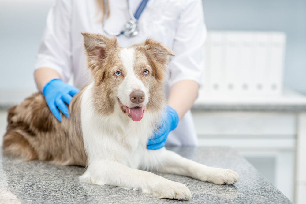
Diagnostics are important to recognize and monitor disease severity and progression. These tests are used as screening tools to detect underlying health issues during routine wellness exams, as well as health complications when a pet is exhibiting signs. Our Sixes Animal Hospital at Bridgemill team shares information about diagnostics that veterinarians commonly use on pets.
Blood work for pets
Blood work is one of the most common diagnostics used to assess a pet’s overall health and detect disease. Common blood work diagnostics include:
- Complete blood count (CBC) — A CBC is used to assess your pet’s white blood cells, red blood cells, and platelets, and can detect conditions such as anemia, infection, blood clotting disorders, and certain cancers.
- Biochemistry profile — A biochemistry profile measures several body systems to detect conditions such as kidney disease, liver disease, electrolyte imbalances, and diabetes.
- Heartworm test — A heartworm test detects specific heartworm proteins (i.e., antigens) that are released by adult female heartworms. These tests can also typically detect tick-borne illnesses.
- Thyroid test — Senior pets are at high risk for thyroid disease, so thyroid panels are used to measure thyroid hormone levels to ensure they are within normal limits.
Cytology for pets
Cytology involves microscopically examining cell samples to determine if abnormalities are present, and to establish a preliminary diagnosis. These samples can be taken from many areas, including the skin, growths or masses on the body surface, internal organs, joints, the eye, and from inside the chest or abdomen. Collection methods that a veterinary professional uses include:
- Fine needle aspiration — A sterile needle attached to an empty syringe is inserted into the collection area, and a sample is aspirated.
- Skin scraping — A needle or scalpel blade is used to scrape the skin and cells are collected on a microscope slide.
- Impression smear — Used most commonly when a pet has an ulcerated skin lesion, a microscope slide is pressed on the affected area and a sample collected.
- Cotton tipped swabs — A cotton swab is used to collect discharge and cells from areas such as the eye, nose, and mouth.
- Lavage — Sterile fluid is flushed into areas, such as the nasal cavity and lungs, suctioned out, and examined.
Histopathology for pets
Histopathology involves microscopically examining whole tissue samples that are collected surgically, preserved, thinly sliced, and stained, and examined by a veterinary pathologist. Histopathology evaluates the tissue architecture, and can provide more information about the tissue than cytology, including a prognosis and a diagnosis for a pet’s condition. Also, a pathologist can use histopathology to determine if a mass was completely removed. Collection methods that a veterinary professional uses include:
- Punch biopsy — A circular blade obtains a circular tissue piece.
- Wedge biopsy — A small slice or chunk is removed from a tumor or mass
- Excisional biopsy — The entire tumor or mass is removed.
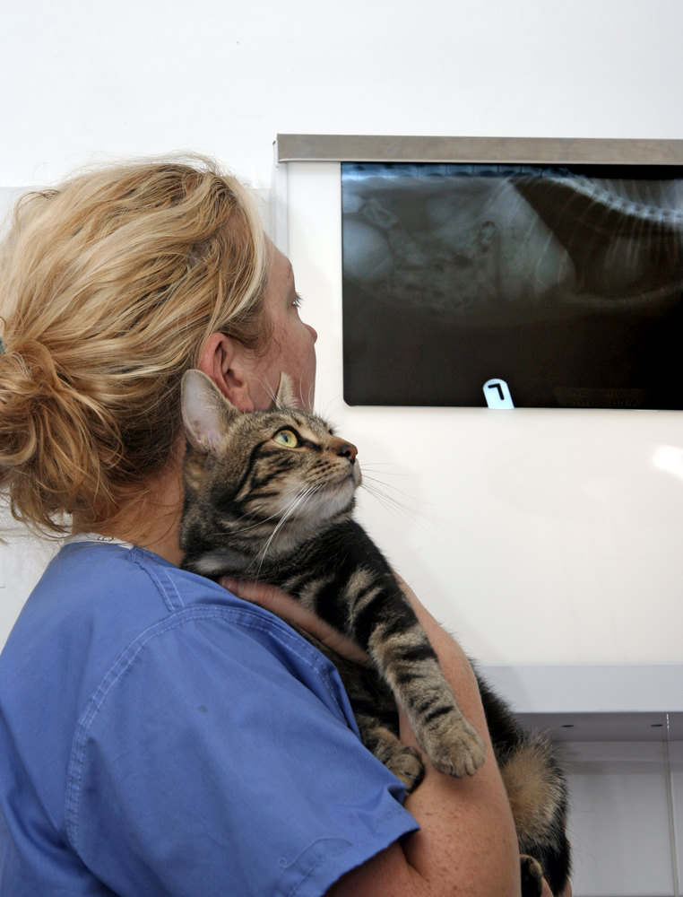
Imaging for pets
Veterinarians use medical imaging to noninvasively view structures inside your pet’s body to help make a diagnosis. The most common imaging techniques include:
- X-rays — An X-ray machine produces a narrow photon beam that can be aimed at a particular body area. As the beam passes through the area, the tissues and bones absorb or block the beam in varying degrees, depending on tissue density, creating an image that is recorded on film or a sensor placed on the other side of the body. On an X-ray, bones appear white, muscles and soft tissues appear gray, and air appears black. X-rays are useful to diagnose issues such as bone fractures, arthritis, pneumonia, heart conditions, and tumors.
- Ultrasound — Ultrasound transducers (i.e., probes) produce sound waves that are sent into the body when the probe is placed on the pet’s skin. When these waves hit boundaries between fluid and tissue or between tissue and bone, they reflect back to the transducer, and generate electrical signals that are sent to the ultrasound scanner. The scanner uses the speed of sound and the time of each reflection’s return to calculate the distance from the transducer to the tissue boundary, producing a two-dimensional image. Ultrasound can be used to examine any body area, including the abdomen, lungs, ligaments, tendons, and eyes.
Advanced imaging for pets
In some cases, pets must be referred for advanced imaging techniques, which include:
- Magnetic resonance imaging (MRI) — MRI creates a strong magnetic field that causes the atoms in the pet’s body to align. The MRI then sends radio waves that stimulate the atoms, which strain against the magnetic field. When the radio waves are turned off, the MRI sensors detect the energy that the atoms release as they realign with the magnetic field. A computer converts the signals into an image. MRI can be used to evaluate the brain and spinal cord, as well as joint abnormalities.
- Computed tomography (CT) — CT uses a motorized X-ray source that rotates around the pet as they slowly move through the CT tube. Each time the X-ray source completes a rotation, the CT computer constructs a two-dimensional image slice of the pet that can be displayed individually, or images can be stacked together to generate a 3D image. CT can be used to evaluate the chest, nasal cavity, joints, spine, and pelvis, and to check for cancer metastasis.
Diagnostics help our veterinary team detect disease and determine why your pet is ill. If your pet is feeling under the weather, contact our Sixes Animal Hospital at Bridgemill team, so we can determine what test is necessary to diagnose the problem.


Leave A Comment